X-ray Diffraction (XRD)
X-ray diffraction (XRD) is an analysis method that directs X-rays at a crystalline sample and measures the directions in which the regular rows of atoms reflect them strongly. Because X-ray wavelengths are similar to atomic spacings, the angles of these reflections reveal the crystal structure, the distance between atomic planes, strain and crystal quality.
Also called: XRD, high-resolution XRD, HRXRD, XRR
Diagram
How diffraction reveals structure
X-rays reflected from successive atomic planes reinforce each other only at angles where their path difference is a whole number of wavelengths. This condition is Bragg's law, nλ = 2d sin θ, where λ is the X-ray wavelength, d is the spacing between planes and θ is the angle of incidence. Measuring diffracted intensity as the angle changes gives a pattern of peaks.
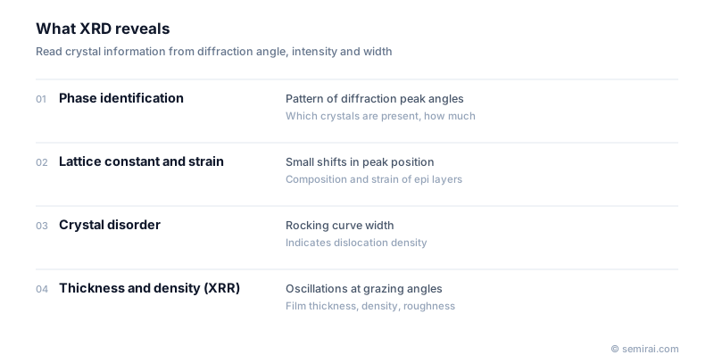
The positions of the peaks identify which crystalline phases are present and give the lattice constants. Small shifts in position reveal strain. The width of the peaks reflects the size of crystal grains and the degree of disorder, and the relative intensities show whether grains share a preferred orientation, called texture.
Uses in semiconductor manufacturing
High-resolution XRD is the standard way to check epitaxial layers. It measures the composition of films such as SiGe, GaN and AlGaN and the strain caused by lattice mismatch with the substrate. The width of a rocking curve, measured by tilting the sample through a reflection, indicates how many dislocations and other defects the crystal contains.
X-ray reflectivity (XRR) uses the same instruments at very shallow angles. Interference between reflections from the top and bottom of a film produces oscillations whose spacing gives thickness and whose shape gives density and interface roughness, even for amorphous films. Because both methods are non-destructive, they are used from research to production monitoring.
Japanese companies involved
- Rigaku makes X-ray diffractometers and X-ray metrology tools, including systems that measure films and epitaxial layers on full wafers.
- Rigaku · X-ray metrology and thermal analysis
FAQ
Can XRD measure amorphous films?
Amorphous films have no long-range order, so they give only broad humps rather than sharp diffraction peaks. Their thickness and density are usually measured by X-ray reflectivity instead, which does not depend on crystallinity.
What X-ray source do laboratory XRD systems use?
Most use a copper anode tube and its Cu Kα line, with a wavelength of about 0.154 nm. High-resolution systems add monochromator crystals to select a very narrow band of wavelengths and angles, which is needed to resolve the small peak shifts from strain in epitaxial layers.
Related pages
- Crystal Structure
- Lattice Constant
- Miller Index
- Epitaxial Wafer (Epi Wafer)
- X-ray Fluorescence (XRF)
- Raman Spectroscopy
- Interplanar Spacing (d-spacing)
- Bragg's Law
- Wafer substrate inspection equipment
- Rigaku FSAS II
- Kobelco Research Institute EPM-2200
- Kobelco Research Institute LEP series
- Kobelco Research Institute SBW series flatness and shape measurement systems
Sources
Last updated: November 13, 2026